NeuroLogic: Fundamentals
EEG Fundamentals
Mastering EEG Fundamentals
Explore the core principles of electrophysiology and master the International 10-20 system for electrode placement. This comprehensive module combines technical theory with interactive practical tools.
How EEG Sees the Brain
When you look at an EEG trace, you aren't seeing individual neurons "firing" action potentials. Instead, you're observing the collective ripple of Postsynaptic Potentials (PSPs).
These PSPs are small shifts in electrical charge that occur when neurotransmitters cross a synapse. Individually, they are microscopic whispers. But when they happen in unison, they become a roar.
Imagine a stadium with 60,000 fans. If they cheer randomly, it's just white noise. But if a Conductor (the Thalamus) coordinates them to cheer in unison, the sound becomes a roar. This is synchronization.
For a signal to reach the scalp, we need roughly 6 square centimeters of cortex to "cheer" at exactly the same time. This massive requirement is due to the insulating properties of the skull and scalp, which act as a low-pass filter, blurring and attenuating high-frequency, asynchronous activity.
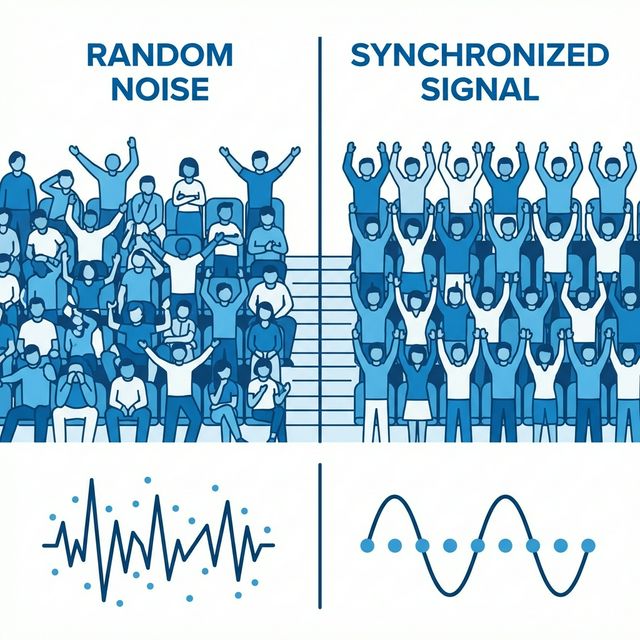
Synchronized populations are visible; individual neurons are too weak alone.
Potentials & Ion Management
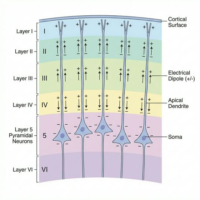
Pyramidal neurons in Layer 5, aligned in parallel, generate the open fields necessary for scalp EEG.
The EEG signal is generated primarily by Pyramidal Neurons arranged in parallel columns within the cortex. These cells act as biological batteries, meticulously managing ions to create electrical fields.
The Dipole Generator
When a neurotransmitter (like Glutamate) binds to the apical dendrites of a pyramidal cell, positive ions (Na+) rush in. This creates a Sink (negative charge) extracellularly at the top, while the cell body becomes a relative Source (positive charge). This separation of charge creates a Dipole.
Action Potentials vs. PSPs: Action potentials are too short (1ms) to summate effectively. PSPs last much longer (10-100ms), allowing them to overlap and build a signal strong enough to reach the scalp.
The Polarity Flip: While an EPSP creates a Negative extracellular field, standard Clinical EEG conventions display Negative values as UP. This is why a negative spike points upward on your screen.
- EPSP (Excitatory): Na+ influx depletes extracellular positive charge, creating a local Negative Sink. Due to the "Negative Up" rule, this creates an Upward Deflection.
- IPSP (Inhibitory): Cl- influx (or K+ efflux) creates a relative Positive Source extracellularly. This creates a Downward Deflection.
Generating the Waveform
The "EEG signal" we read is the sum of these extracellular charge shifts. Because pyramidal neurons are arranged in neat columns perpendicular to the brain's surface, their electrical fields summate linearly. If they were randomly oriented, their fields would cancel each other out (Closed Field).
| Event Location | Charge at Surface | EEG Deflection |
|---|---|---|
| Superficial EPSP (Excitation) | Negative (-) | Upgoing |
| Superficial IPSP (Inhibition) | Positive (+) | Downgoing |
The Universal Rule: In clinical EEG, Negative is UP and Positive is DOWN. While initially counterintuitive, this convention is the standard across all modern neurophysiology.
Orientation & Detection
Even a massive signal can "blind" the EEG if it's pointed the wrong way. This is the concept of Dipole Orientation.
Think of an electrical dipole like a flashlight beam. A "Radial" dipole on top of a gyrus shines directly at the skull (Perpendicular), creating a broad, strong field. A "Tangential" dipole in a sulcus shines sideways, parallel to the skull.
Tangential dipoles are notoriously deceptive. They can create a "paradoxical" field where the dipole projects to the opposite hemisphere, or they may be completely invisible to scalp electrodes directly above them (the "blind spot" of EEG).
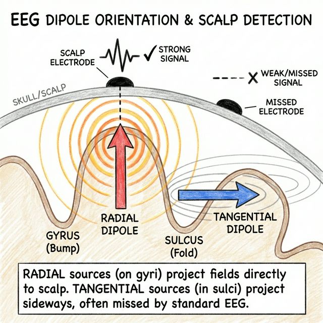
Radial sources (Gyri) are easily seen. Tangential sources (Sulci) project sideways or cancel out.
Mastery Checklist
EEG detects Postsynaptic Potentials (PSPs), not Action Potentials.
Signal visibility requires 6cm² of synchronized cortical area.
Open Fields: Parallel alignment of Pyramidal cells is critical for summation.
In EEG interpretation: Up is Negative, Down is Positive.
Tangential Dipoles (in sulci) are harder to localize than Radial Dipoles.
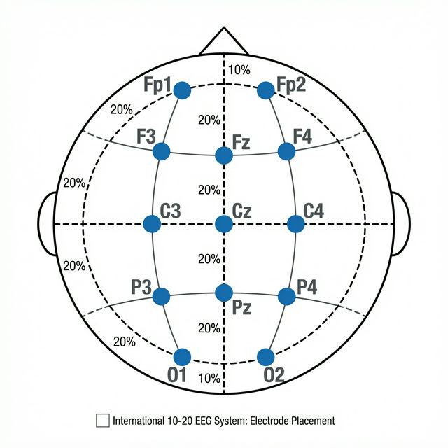
The 10-20 System: A Global Map
Consistency is key. The International 10-20 System uses percentages (10% and 20%) of skull distances to place electrodes. This ensures that "C3" is always over the primary motor cortex, regardless of head size.
- Letters: F (Frontal), T (Temporal), C (Central), P (Parietal), O (Occipital).
- Numbers: Odd (Left), Even (Right), z (Midline).
Side View (Profile)
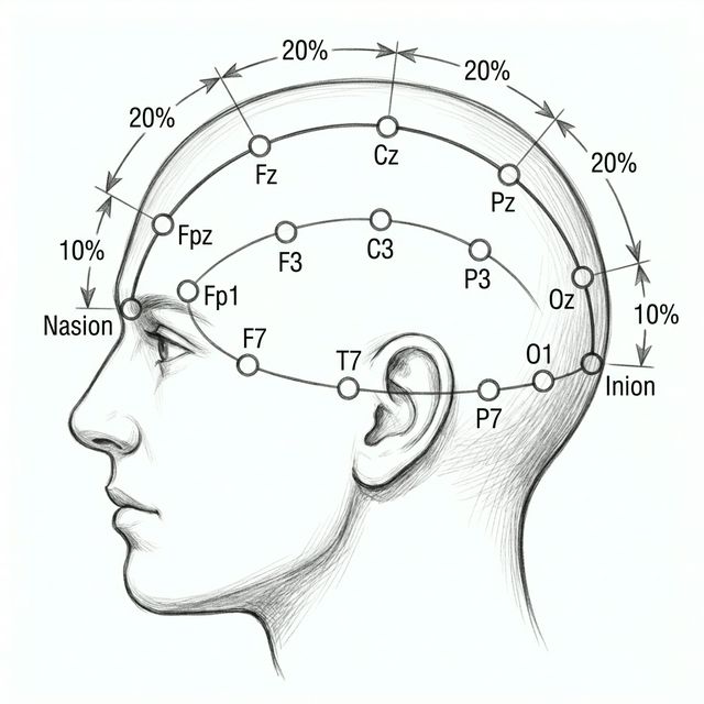
Top View
The Grid: Understanding Montages
An electrode on the scalp is just a piece of metal. It has no value until it is compared to something else. This comparison (Input 1 minus Input 2) is the heart of differential amplification.
An EEG Montage is a logical arrangement of these comparisons. The most common standard is the "Double Banana" (Longitudinal Bipolar) montage, which arranges chains from front-to-back over the left and right hemispheres.
Common Mode Rejection (CMR): The amplifier rejects signals that are identical at both inputs (like 60Hz hum affecting the whole head equally) and amplifies only the difference between them.
Bipolar Logic: The Daisy Chain
In a Bipolar Montage, we link adjacent electrodes in a chain (e.g., Fp1-F3, F3-C3, C3-P3). Each channel compares a specific point to its immediate neighbor.
Channel = Input 1 - Input 2
If Input 1 is -50μV and Input 2 is -20μV:
(-50) - (-20) = -30μV (Upward Deflection).
The Phase Reversal
This is the "Holy Grail" of bipolar localization. When a negative discharge is located directly under an electrode (e.g., F3), it becomes the most negative point in the chain.
- Channel 1 (Fp1-F3): F3 is Input 2. Input 2 is very negative. Result: Positive (Down).
- Channel 2 (F3-C3): F3 is Input 1. Input 1 is very negative. Result: Negative (Up).
The waves point toward each other, "kissing" at the point of maximum negativity.
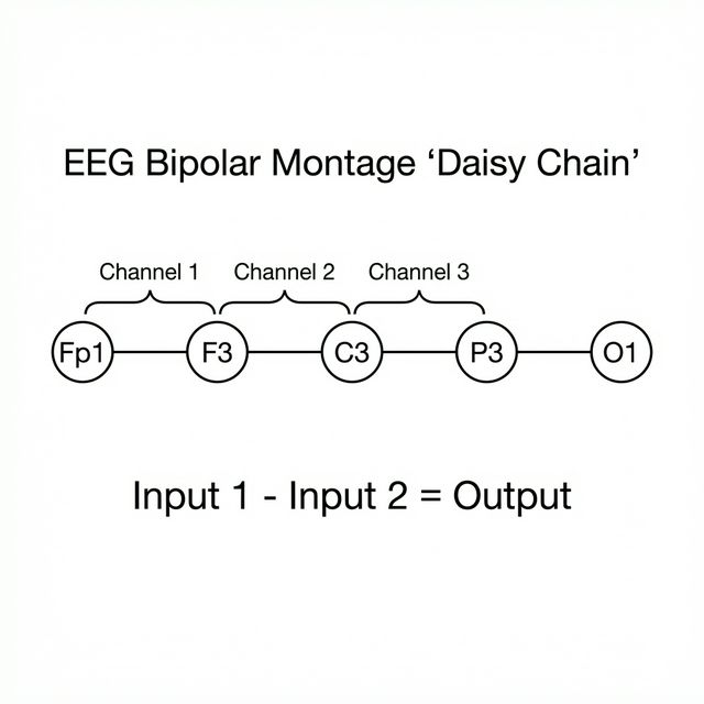
Bipolar chains create a localized view, excellent for finding focal spikes via phase reversals.
Scalp View
Channel 1: Fp1 - F3
Channel 2: F3 - C3
Referential Power: The Benchmarks
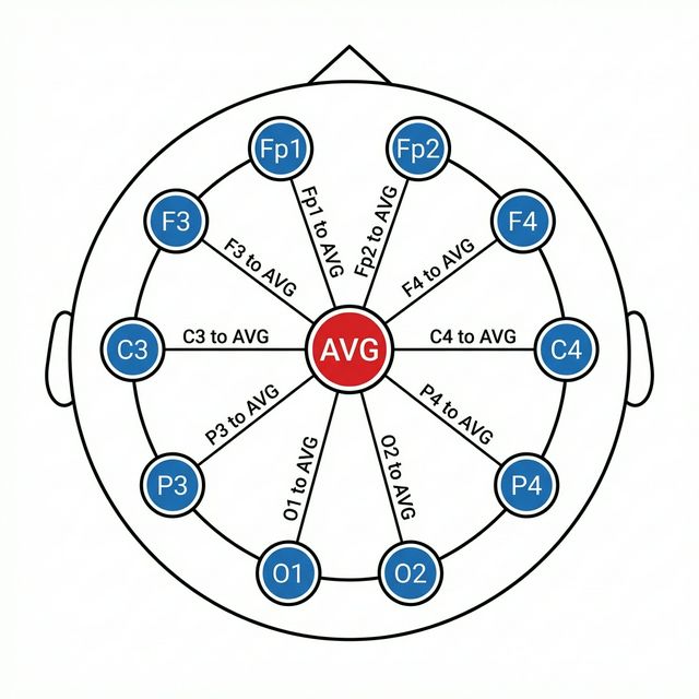
All channels compare to a calculated average of all electrodes (CAR). Amplitude indicates proximity to the source.
Common Average Reference (CAR): This montage calculates the average potential of all scalp electrodes and uses this computed signal as the reference for each channel. It helps eliminate common, widespread signals in favor of electrode-specific activity, making it a versatile, non-biased screening tool.
Amplitude = Proximity
The channel with the highest amplitude is closest to the source. No phase reversals here.
Contaminated Reference
It is susceptible to "reference contamination" from high-voltage artifacts, which can distort the data.
Control Panel: Filters & Sensitivity
LOWER NUMBER = HIGHER ZOOM
At 7uV/mm, a 70uV wave = 10mm tall.
Setup Essentials Checklist
Bipolar Montages use phase reversals ("kissing waves") to localize.
Referential Montages use maximum amplitude to localize.
Common Mode Rejection eliminates noise present at both inputs.
Time Constant is the inverse of Frequency for LFF settings.
EEG: A Language of its Own
Once you've got the electrodes in place, montage chosen, and the study started, it's your job to interpret and communicate the EEG findings. The first step is learning how to differentiate the many waveforms you'll come across, and the vocabulary you need to communicate what you see.
Think of waveforms as the "words" of EEG. Just as words have length and sound, waveforms have Frequency and Amplitude.
Frequency: The Speed of Thought
Recognizing the frequency of the waveforms is fundamental to interpreting EEG. Frequency describes how many waves there are per second, and is measured in Hertz (Hz).
There are four main frequency bands in the human brain seen on scalp EEG, in increasing order: Delta, Theta, Alpha, and Beta.
Delta (0-4 Hz)
The slowest rhythm. Hallmark of deep sleep; otherwise points to structural issues or encephalopathy.
Theta (4-8 Hz)
Common in children and during drowsiness. Hallmark of normal "RMTD" findings.
Alpha (8-13 Hz)
The "Gold Standard" of the awake adult brain. Forms the Posterior Dominant Rhythm (PDR), which appears in the occipital regions when eyes are closed and disappears when eyes open (reactivity).
Beta (13-30 Hz)
Fast activity often seen with muscle artifact or benzodiazepine use. Gamma (>30Hz) is rarely seen on scalp.
Note: Real EEG is rarely "pure." You'll often see delta with overriding beta—a mixture of slow and fast activity happening at once.
Visualizing Frequencies
Delta: 0.5-4 Hz (Slow & High Amplitude)
Theta: 4-8 Hz (Intermediate)
Alpha: 8-13 Hz (Normal Awake Rhythm)
Beta: 13-30 Hz (Fast & Low Amplitude)
Universal Law: Physiologic waves usually have an inverse relationship between frequency and amplitude. Lower frequency (Delta) = Higher Amplitude; Higher frequency (Beta) = Lower Amplitude.
When the Brain Sleeps
Sleep isn't just "off." It is a complex, staged architecture with unique signatures.
- N1 (Drowsiness): The PDR (Alpha) drops out. You see Slow Rolling Eye Movements.
- N2 (Light Sleep): The hallmark stage. Look for Sleep Spindles (fast 14Hz bursts) and K-Complexes (massive, biphasic dips).
- N3 (Deep Sleep): Dominated by high-amplitude, slow Delta waves.
- REM: "Paradoxical Sleep." The EEG looks almost awake (mixed frequency), but the patient is paralyzed (atonia) and dreaming.
Amplitude: The Power of the Wave
Amplitude is the height of a waveform—a proxy for voltage. In scalp EEG, this is measured in microvolts (μV).
A normal adult brain typically produces signals between 10 to 100 μV, with most clinical activity falling in the 10-50 μV range.
Don't Confuse Amplitude and Sensitivity: Amplitude is an intrinsic trait of the brain signal. Sensitivity is a technical dial (like 7μV/mm) that you adjust to see the signal better.
Morphology & Rhythmicity
Morphology describes the overall shape of a waveform. To break it down, we look at Phases (how many times a wave crosses the baseline).
- Monophasic: Stays on one side of the baseline. Example: A simple clinical spike.
- Biphasic: Crosses the baseline once. Example: The classic epileptiform spike and slow wave discharge.
- Polyphasic: Crosses the baseline multiple times.
The Pattern of the Trace:
Monomorphic (Rhythmic)
Waves that look identical in frequency and amplitude. Often concerning for epileptiform activity.
Polymorphic
Waves that vary constantly. A hallmark of slow-wave sleep or focal slowing from tumors/bleeds.
Artifacts: The Real World Noise
Not everything on the EEG comes from the brain. You must learn to filter out the noise.
Eye Blinks
The eye is a battery (Cornea +, Retina -). Blinking creates massive "Frontal Dipoles" seen clearly in Fp1/Fp2.
Muscle (EMG)
High-frequency buzzing/blacking out of channels. Common in Temporals from jaw clenching.
60Hz Hum
Electrical interference from the wall. Looks like a thick black line. Removed by the Notch Filter.
The Waveform Dictionary
Frequencies: Delta (0-4Hz), Theta (4-8Hz), Alpha (8-13Hz), Beta (13-30Hz).
Amplitude: Measures voltage in microvolts (Normal: 10-100μV).
Morphology: Shape classified by phases (Monophasic, Biphasic, Polyphasic).
Patterns: Monomorphic (repetitive/rhythmic) vs Polymorphic (varying).
Available Leads
Results
Lead Placement Quiz
Click "Start Quiz" to begin testing your knowledge of lead placement.